Malaria? Be confident.
Malaria, for some, may present as a faraway tropical disease, but for others is indeed an everyday challenge. Travelling and migration though have brought malaria to the doorsteps of virtually every country on the planet. Sysmex, identifying this threat and building on its haematology expertise, can help you identify with confidence a malaria infection, guiding treatment decisions and evaluating treatment success, no matter if you are dealing with malaria on an everyday basis, or you occasionally meet travellers suspected of an infection.
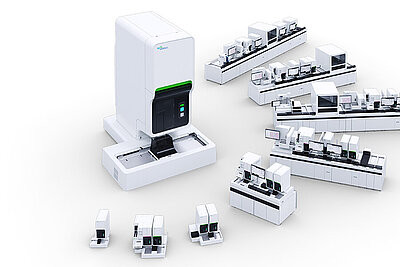
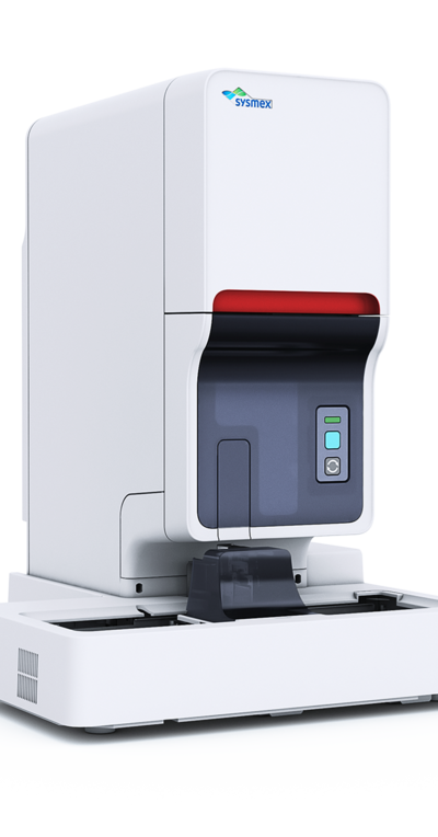
On this page you can explore the benefits from Sysmex’s solution to malaria diagnosis, the XN-31 analyser, see XN-31 in action using real-life examples and read about the recommended diagnostic guidelines from the World Health Organisation (WHO) and other medical associations all over the world.
Benefits
- Sensitive detection and count of malaria-infected red blood cells (MI-RBC) within just one minute.
- All relevant information for treatment decision in one analysis: parasitaemia load and the suspected parasite species.
- A concurrent complete blood count assists in determining other clinical conditions, like anaemia.
- Reliable results for the whole course of the disease with a fully standardised analysis: from diagnosis to therapy monitoring.
Simultaneous diagnosis of malaria and anaemia in a paediatric sample
An infant under the age of 1 year old was examined in a hospital of the malaria endemic country Burkina Faso. Blood analysis on the XN-31 analyser identified malaria-infected red blood cells with a high parasitaemia load (MI-RBCa = 68.09x10³ cells/μL) in combination with the presence of mostly ring forms, thus a diagnosis of malaria was made, probably caused by Plasmodium falciparum. Further information from the complete blood count on the same analyser revealed the presence of microcytic hypochromic anaemia (MCV = 50.9 fL, MCHC = 25.9 g/dL and HGB = 5.6 g/dL). Based on these findings, a severe malaria was diagnosed, and a single intravenous treatment with 45 mg artesunate was initiated, as recommended by the WHO. A follow-up two days later revealed the successful course of the treatment with a significant reduction in the parasitaemia load (MI-RBC = 0.21x10³ cells/μL).
A fast and reliable diagnosis of a malaria infection with anaemia was conducted using a single analyser that does not require the specialised training of microscopy. Detailed red blood cell indices showed that a severe malaria could be underway, thus a specialised treatment was immediately initiated, that proved to be successful. Distinguishing between a severe and an uncomplicated malaria is important for choosing the correct therapeutic regimen. An uncomplicated malaria is treated differently than a severe malaria case described above. One of the recommended artemisin-based combination therapies for uncomplicated malaria is given orally for 3 days, in order to clear the parasites from the blood as soon as possible and its progression to a severe case.
a. MI-RBC: malaria-infected red blood cells
Fast and confident treatment monitoring in an adult traveller
A 45-year old male, permanent resident of a malaria non-endemic European country, visited Tanzania for a two-week holiday. Three days after returning in his home country, he started to have fever, shivers and a headache. Five days later, when symptoms had worsened and by then also included myalgia, arthralgia and malaise, he visited the local hospital. When the patient presented at the emergency department, he had a fever of 39° Celsius, weakness and reduced awareness. As the patient was severely ill and recently returned from tropical areas, malaria was suspected. Hence, the treating physician ordered malaria examination and a panel of other laboratory examinations. (see table below)
A rapid diagnostic test (RDT) demonstrated the presence of HRP-2, an antigen specific for Plasmodium falciparum. Detailed blood analysis on the XN-31 analyser revealed an ongoing malaria infection, probably caused by P. falciparum, with a very high parasitaemia load (MI-RBCa = 539 x 103 cells/μL) and the presence of trophozoites (ring forms) and schizonts. These results demonstrated that the patient suffered from severe malaria, thus he was admitted to the intensive care unit (ICU) and therapy was initiated immediately. As per hospital guidelines, the results were confirmed by a quantitative buffy coat (QBC) and a microscopic examination of thin and thick blood smears.
The patient was initially treated with intravenous artesunate (2.4 mg/kg at 0, 12 and 24 hours after diagnosis), after which treatment was continued with oral administration of atovaquone/proguanil combination (1dd 1000/400 mg for 3 days). During the treatment period, parasitaemia was determined regularly with the XN-31 analyser until parasites were cleared. The patient was discharged from the hospital after 7 days and subsequently fully recovered.
The presence of an analyser that combines the speed of an RDT with the parasite quantification features of a microscopic examination, and does not require an expert user was instrumental for rapidly diagnosing malaria outside office hours and acting fast on treatment initiation. Moreover, the high sensitivity of the analyser in detection of malaria-infected RBC (down to 20 per microliter of blood) was useful in the follow-up measurements during treatment and provided confidence in deeming the treatment successful.
a. MI-RBC: malaria-infected red blood cells
Table with laboratory examinations upon hospital admission
Clinical condition | Examination | Result |
| Hypoglycaemia | Glucose | 3.9 mmol/L |
| Anaemia | Haemoglobin | 7.3 mmol/L |
| Thrombocytopenia | Platelet count | 16x103 cells/μL |
| Renal dysfunction | Urea Creatinine Glomerular filtration rate | 10.8 mmol/L 115 μmol/L 49 mL/min |
| Disturbed plasma electrolytes | Sodium Potassium Calcium | 131 mmol/L 3.8 mmol/L 2.08 mmol/L |
| Acidosis | Plasma lactate | 5.4 mmol/L |
| Bilirubinaemia | Total plasma bilirubin | 119 μmol/L |
| Organ damage | ALAT ASAT γGT Alkaline phosphatase Lactate dehydrogenase | 164 U/L 174 U/L 168 U/L 101 U/L 1015 U/L |
| Inflammation | C-reactive protein (CRP) | 181 mg/L |
| Haemocytometric profile | White blood cells Monocytes Neutrophils Plasma cells | 12.7% 3.5% 38.1% 4.1% |
It’s not always COVID-19
The COVID-19 pandemic has shifted the focus from other killer diseases in several aspects, such as diagnostics and public awareness. The situation is very critical in the fight against malaria, where years of efforts could be lost in a very short time. Indeed, for the first time since 2016, the number of malaria deaths in 2020 increased compared to the year before. Approximately 36,000 additional deaths can be attributed to disruptions in health services brought by the COVID-19 pandemic.
Cases of malaria and SARS-CoV-2 co-infections that go unnoticed or missed malaria diagnoses* due to the focus on COVID-19 highlight the importance of being vigilant for all possible aetiologies. Patients with overlapping symptoms must be identified, travel history must always be considered in the differential diagnosis of febrile patients [1, 2, 3]. In that direction, the WHO urges the medical community to continue prevent and treat killer diseases, such as malaria [4].
[1] Sardar et al., IDCases (2020) e00879 | [2] Normal et al., Journal of Travel Medicine (2020) 1–2 | [3] Jochum et al., Trop. Med. Infect. Dis. (2021) 6, 40 | [4] WHO Malaria report 2021
International guidelines for the diagnosis of malaria
Medical associations and ministries of health all over the world, in line with the WHO recommendations, consider the microscopic examination of thick and thin blood films as the gold standard for the detection of malaria parasites. A thick blood film is used to detect malaria parasites, and upon positivity, the thin blood film can support the identification of the Plasmodium species.
Developed for a quick identification of malaria parasites in endemic countries, where laboratory workload is very high, rapid diagnostic tests (RDT) can also be used as a parasitological test to confirm the diagnosis, as per the WHO guidelines. RDTs were developed and are mainly used for the detection of suspected falciparum infections, but newer generation of tests with pan-specific antigen targets, have also been shown to be effective for the non-falciparum malaria species.
Medical guidelines in endemic African countries require the use of microscopy where it is applicable, mainly in well-equipped tertiary health institutions. For primary or secondary care, an RDT can be the first diagnostic tool, but only as an alternative or complementary to microscopy. Species determination and parasite quantification can only be done with microscopic examination.
Medical guidelines in non-endemic countries dictate that a positive RDT must always be followed up by microscopical examination. UK guidelines for example, consider RDTs as an initial screen when laboratory expertise is not available, but they should not be considered as a replacement for blood film examinations.
Molecular techniques such as PCR are not part of the WHO guidelines, due to insufficient standardisation and validation, but local guidelines in non-endemic countries reserve a role for PCR for the identification of Plasmodium species in the highly specialised reference laboratories.
Guidelines and recommendations
- WHO: Guidelines for the treatment of malaria
- UK malaria treatment guidelines
- Guidelines for malaria case management in Ghana
- Guidelines for the treatment of malaria in Malawi
- Canadian recommendation for the prevention and treatment of malaria
- National policy on malaria diagnosis and treatment in Nigeria
Informative sources
- WHO: World malaria report 2020
- WHO: Malaria in the European region
- Malaria factsheet from the European Centre for Disease Prevention and Control
- Malaria diagnosis in the United States from the Center for Diseases Control
- The laboratory diagnosis of malaria in the UK
- Management of imported malaria in Europe


