Scientific Image Gallery
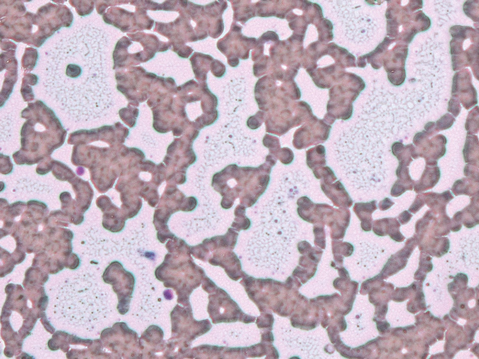
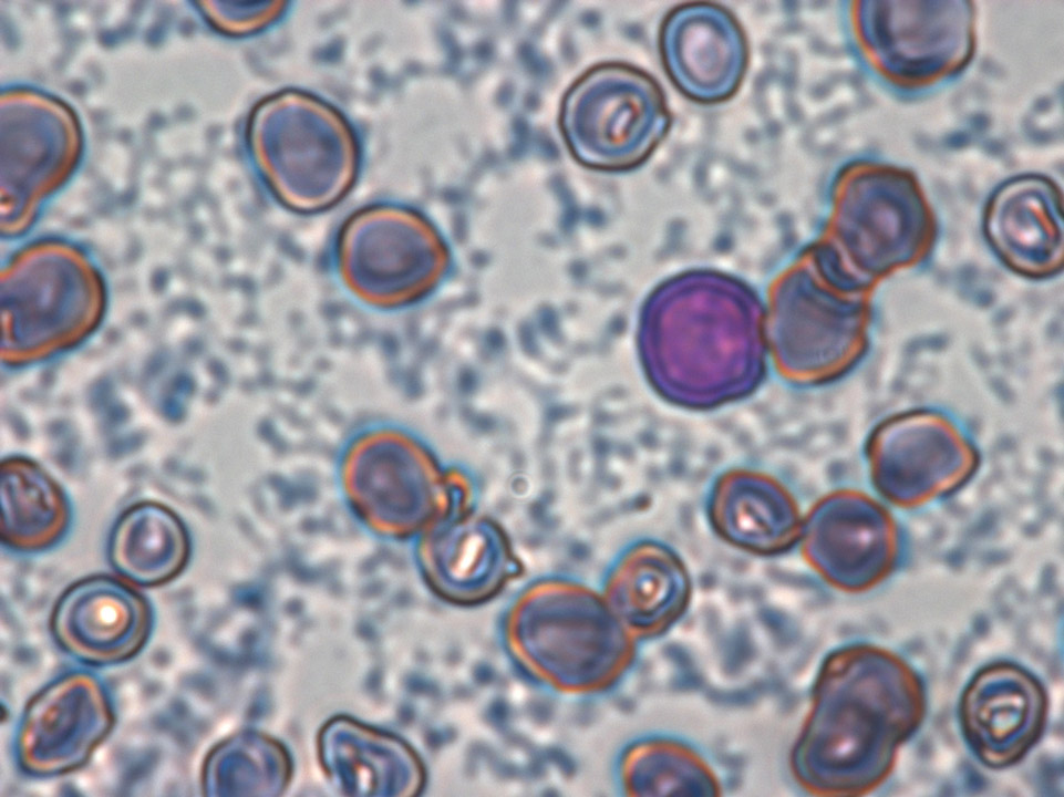
In the impedance channel of a haematology analyser a falsely elevated platelet concentration of 850,000/μL was measured. This was caused by precipitated cryoglobulin in cryoglobulinaemia. The flocculated cryoglobulin is visible in the phase contrast microscope in between the red blood cell 'rouleaux'.
<p>In the impedance channel of a haematology analyser a falsely elevated platelet concentration of 850,000/μL was measured. This was caused by precipitated cryoglobulin in cryoglobulinaemia. The flocculated cryoglobulin is visible in the phase contrast microscope in between the red blood cell 'rouleaux'.</p>
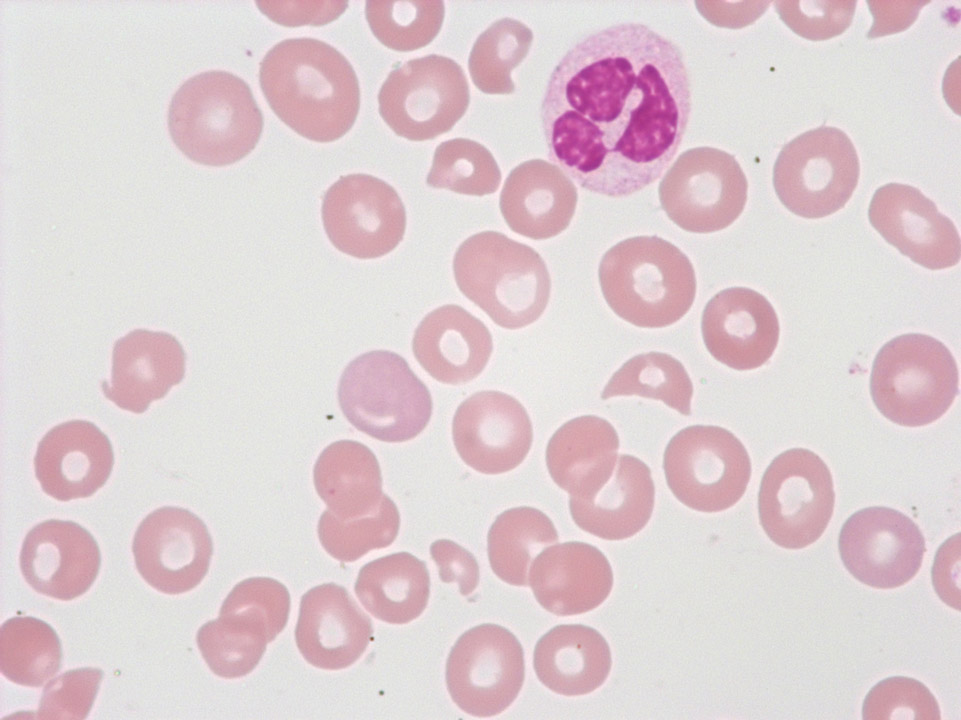
Fragmented red blood cells and thrombocytopenia in the case of a thrombotic thrombocytopenic purpura (TTP). In the partially thrombosed capillaries the red blood cells are exposed to a high degree of shearing force, which causes them to burst.
<p>Fragmented red blood cells and thrombocytopenia in the case of a thrombotic thrombocytopenic purpura (TTP). In the partially thrombosed capillaries the red blood cells are exposed to a high degree of shearing force, which causes them to burst.</p>
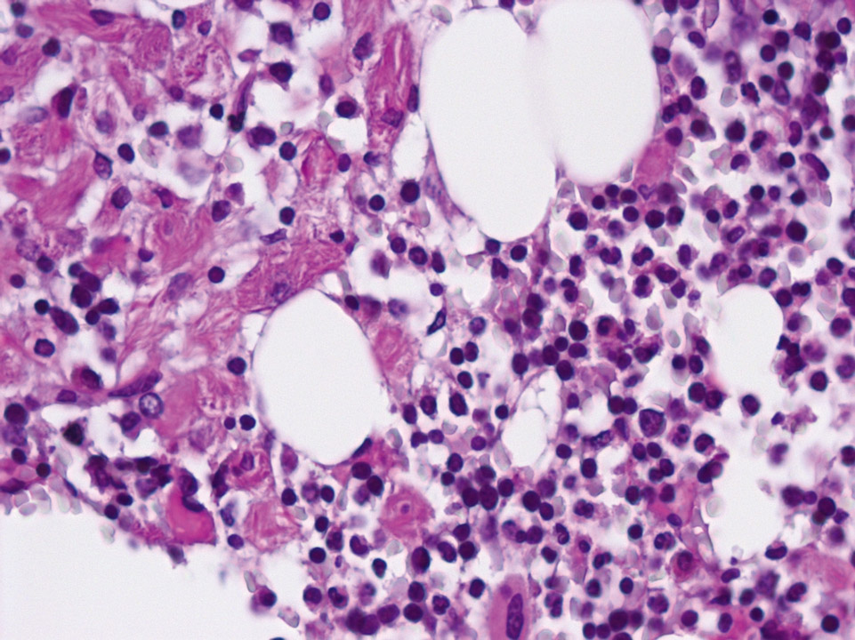
Bone marrow histology (periodic acid Schiff stain, PAS) displaying macrophages on the left, with a cytoplasm resembling crinkled parchment. They are so-called 'Gaucher' cells. Gaucher's disease is characterised by a congenital defect of the enzyme glucocerebrosidase leading to an increase of certain glycolipids (cerebrosides) inside the macrophages. On the right side of the picture the bone marrow appears normal.
<p>Bone marrow histology (periodic acid Schiff stain, PAS) displaying macrophages on the left, with a cytoplasm resembling crinkled parchment. They are so-called 'Gaucher' cells. Gaucher's disease is characterised by a congenital defect of the enzyme glucocerebrosidase leading to an increase of certain glycolipids (cerebrosides) inside the macrophages. On the right side of the picture the bone marrow appears normal.</p>
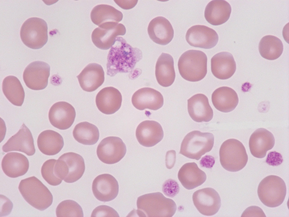
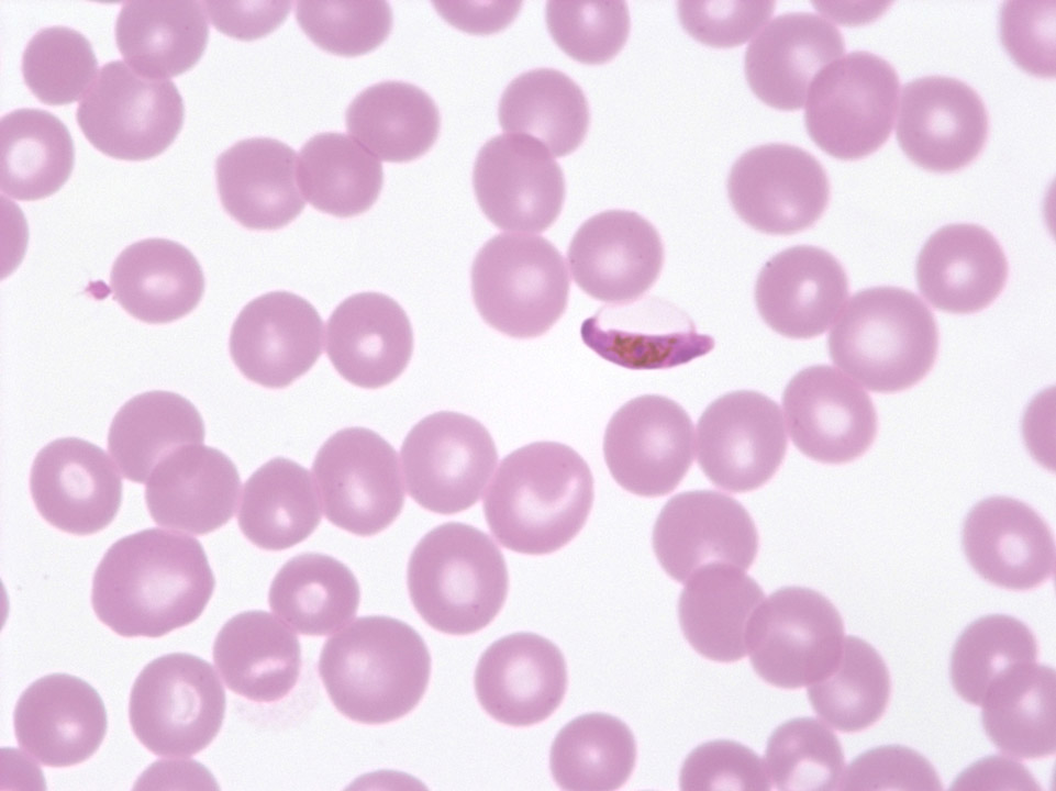
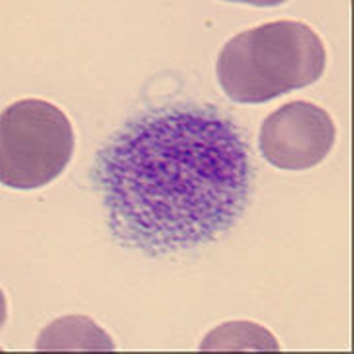
Giant platelet with prominent granulation from a patient with essential thrombocythaemia (ET). (Defective haematopoiesis causes detectable poikilocytosis.)
<p>Giant platelet with prominent granulation from a patient with essential thrombocythaemia (ET). (Defective haematopoiesis causes detectable poikilocytosis.)</p>
