Scientific Image Gallery
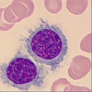
Cell description:
Size: larger than normal lymphocytes
Nucleus: round, oval, dumbbell-shaped or bilobed with little chromatin condensation and sometimes indistinct nucleolus
Cytoplasm: abundant weakly basophilic with irregular “hairy” margins
<p>Cell description: </p> <p>Size: larger than normal lymphocytes </p> <p>Nucleus: round, oval, dumbbell-shaped or bilobed with little chromatin condensation and sometimes indistinct nucleolus </p> <p>Cytoplasm: abundant weakly basophilic with irregular “hairy” margins</p>
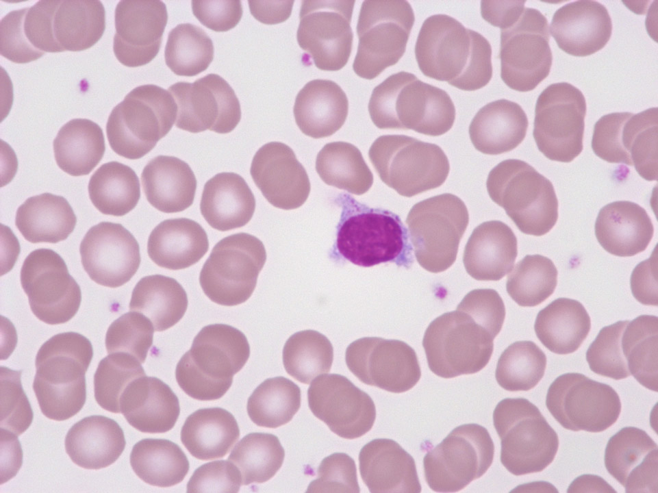
This 'hairy' lymphocyte comes from a person suffering from an active infection (without any malignant disease). The granula identify it as a T or an NK cell.
<p>This 'hairy' lymphocyte comes from a person suffering from an active infection (without any malignant disease). The granula identify it as a T or an NK cell.</p>
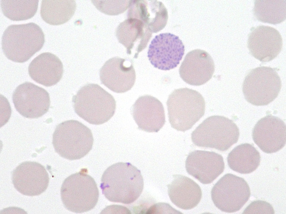
Reticulocyte staining. Characteristic HbH cell in a case of α-thalassaemia. Today, blood films are no longer investigated for HbH cells. Instead,
α-thalassaemias are diagnosed by molecular genetic tests.
<p>Reticulocyte staining. Characteristic HbH cell in a case of α-thalassaemia. Today, blood films are no longer investigated for HbH cells. Instead, </p> <p>α-thalassaemias are diagnosed by molecular genetic tests.</p>
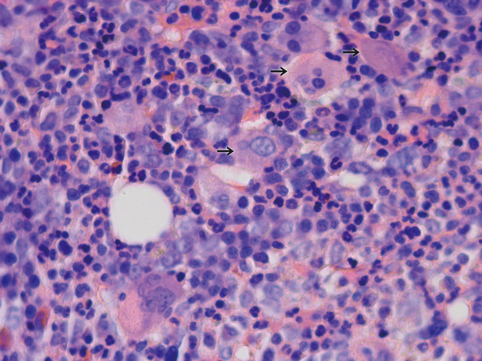
A remarkably high and conspicuous number of megakaryocytes (->) is visible in the bone marrow histology (Giemsa stain) of a patient with CML.
<p>A remarkably high and conspicuous number of megakaryocytes (->) is visible in the bone marrow histology (Giemsa stain) of a patient with CML. </p>
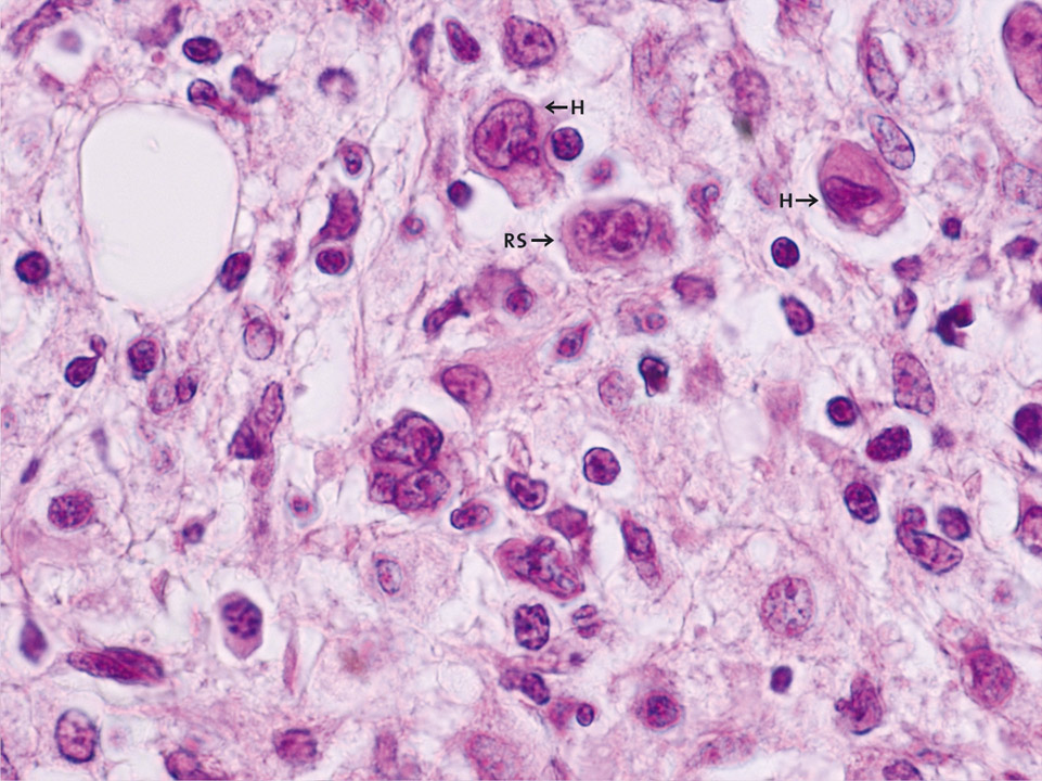
Bone marrow histology (Giemsa stain) showing two Hodgkin cells (H). These giant cells have an oval, non-lobulated nucleus and at least one nucleolus. Reed-Sternberg cells (RS) have a lobulated nucleus with several nucleoli. Both cell types are characteristic of Hodgkin's disease and express the same surface marker CD30, which is otherwise found on activated lymphocytes.
<p>Bone marrow histology (Giemsa stain) showing two Hodgkin cells (H). These giant cells have an oval, non-lobulated nucleus and at least one nucleolus. Reed-Sternberg cells (RS) have a lobulated nucleus with several nucleoli. Both cell types are characteristic of Hodgkin's disease and express the same surface marker CD30, which is otherwise found on activated lymphocytes. </p>
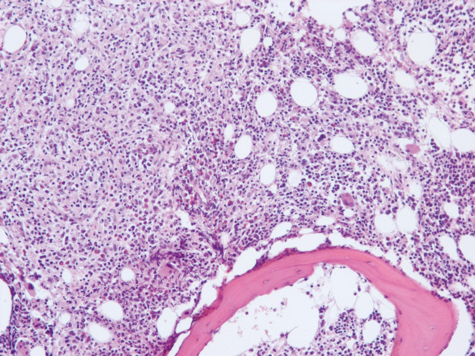
Bone marrow histology (Giemsa stain) of a patient showing an infiltrating Hodgkin's lymphoma on the left side. On the right side normal haematopoietic tissue can be seen.
<p>Bone marrow histology (Giemsa stain) of a patient showing an infiltrating Hodgkin's lymphoma on the left side. On the right side normal haematopoietic tissue can be seen.</p>
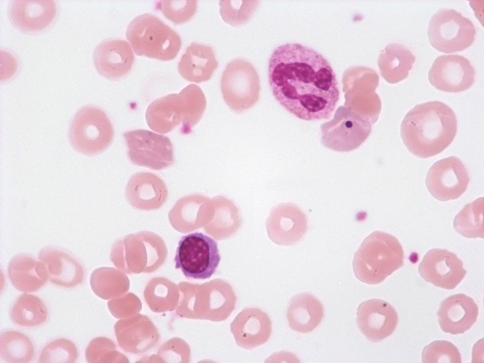
Howell-Jolly bodies in the cell adjacent to the granulocyte, spherocytes, polychromasia, and a single erythroblast in a case of auto-immune haemolytic anaemia (AIHA). (Howell-Jolly bodies are DNA remnants. Polychromatic cells still contain RNA.)
<p>Howell-Jolly bodies in the cell adjacent to the granulocyte, spherocytes, polychromasia, and a single erythroblast in a case of auto-immune haemolytic anaemia (AIHA). (Howell-Jolly bodies are DNA remnants. Polychromatic cells still contain RNA.)</p>
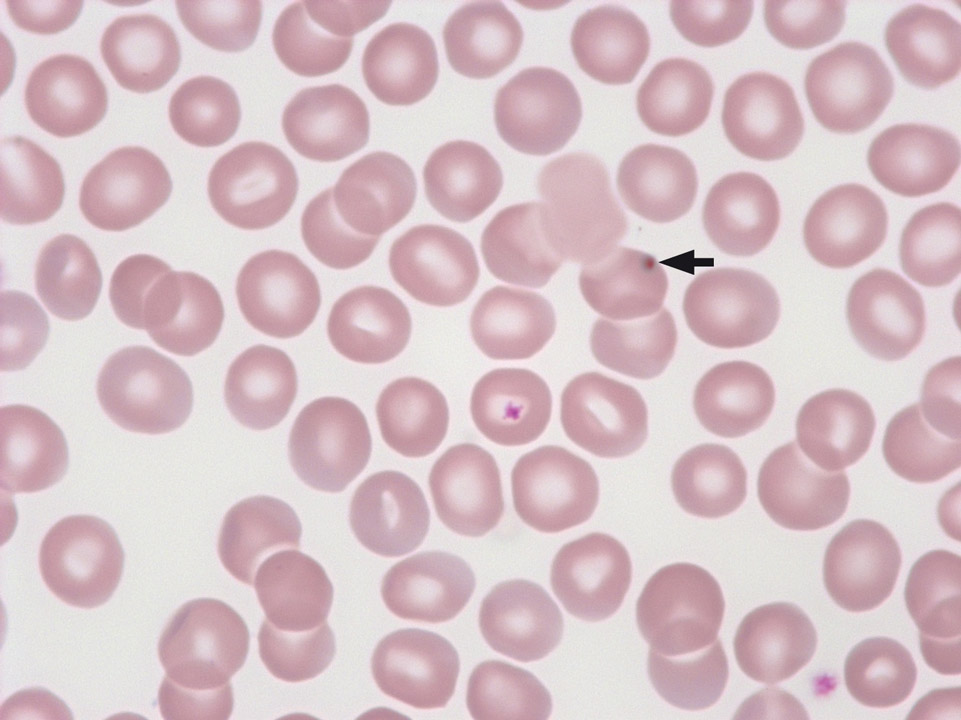
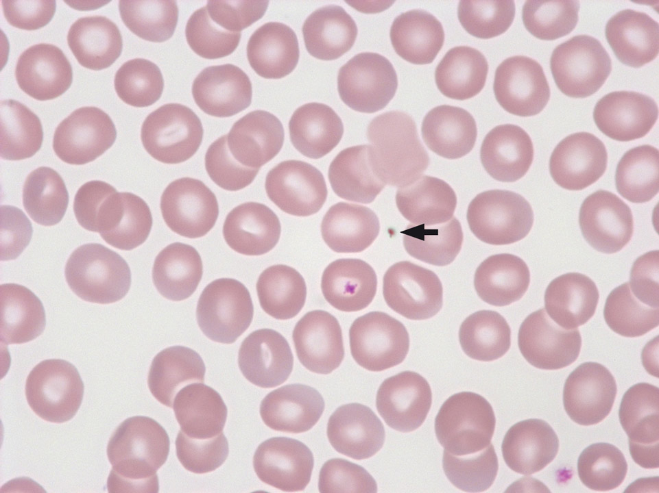
An apparent Howell-Jolly body, caused by contaminated microscope oil, can be identified as an artefact by the fact that it moves independently over time.
<p>An apparent Howell-Jolly body, caused by contaminated microscope oil, can be identified as an artefact by the fact that it moves independently over time.</p>