Scientific Image Gallery
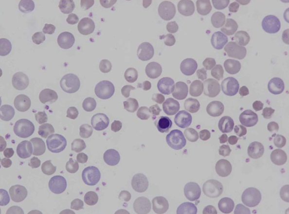
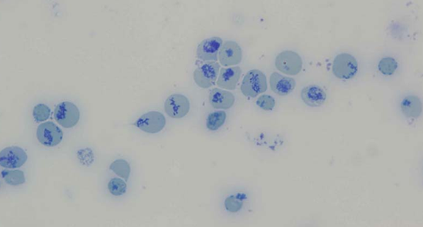
Blood smear of a rat showing marked anisocytosis, poikilocytosis and polychromasia. A nucleated red blood cell can be seen in the centre.
<p>Blood smear of a rat showing marked anisocytosis, poikilocytosis and polychromasia. A nucleated red blood cell can be seen in the centre.</p>
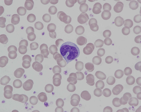
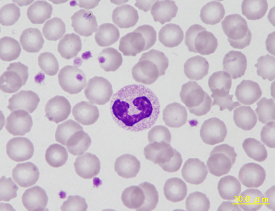
Typical monocyte of a rat. Both the colour shade of the cytoplasm and the shape of the nucleus reflect distinctive characteristics of this cell type.
<p>Typical monocyte of a rat. Both the colour shade of the cytoplasm and the shape of the nucleus reflect distinctive characteristics of this cell type.</p>
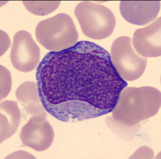
The first microscopically identifiable cell of granulocytic cell line.
Cell description:
Size: 12-20 µm
Nucleus: large, round or slightly oval with diffuse chromatin pattern and often 1-5 nucleoli
Cytoplasm: pale blue and usually agranular, sometimes Auer rods visible
<p>The first microscopically identifiable cell of granulocytic cell line. </p> <p>Cell description: </p> <p>Size: 12-20 µm </p> <p>Nucleus: large, round or slightly oval with diffuse chromatin pattern and often 1-5 nucleoli </p> <p>Cytoplasm: pale blue and usually agranular, sometimes Auer rods visible</p>
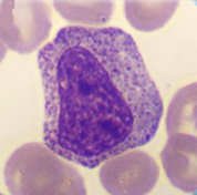
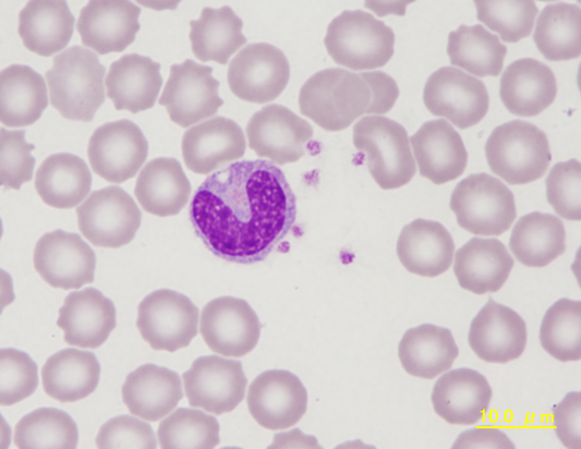
In this maturation stage the separation into the 3 different subpopulations of granulocytes occurs by development of specific granulation for each (secondary granulation).
Cell description:
Size: 10-18 µm
Nucleus: oval or slightly indented with variable degree of chromatin clumping, nucleoli usually not apparent
Cytoplasm: acidophilic neutrophil: primary azurophilic and secondary neutrophilic granules
<p>In this maturation stage the separation into the 3 different subpopulations of granulocytes occurs by development of specific granulation for each (secondary granulation). </p> <p>Cell description: </p> <p>Size: 10-18 µm </p> <p>Nucleus: oval or slightly indented with variable degree of chromatin clumping, nucleoli usually not apparent </p> <p>Cytoplasm: acidophilic neutrophil: primary azurophilic and secondary neutrophilic granules</p>
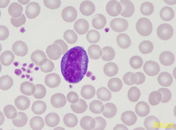
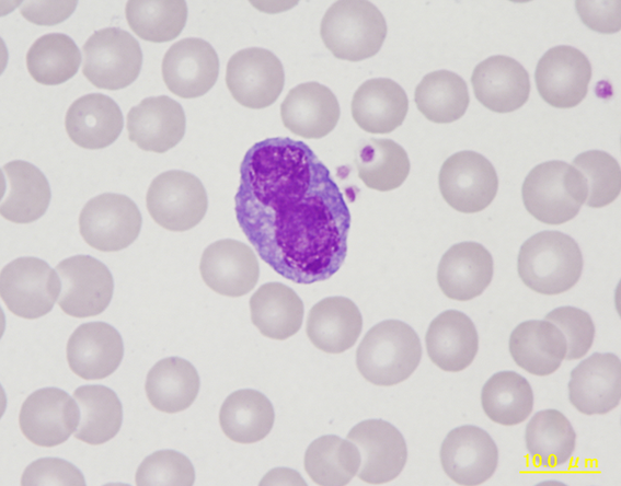
Atypical lymphocyte with a large in size and intensely blue staining cytoplasm. Atypical lymphocytes are defined as lymphocytes responding to antigenic stimuli.
<p>Atypical lymphocyte with a large in size and intensely blue staining cytoplasm. Atypical lymphocytes are defined as lymphocytes responding to antigenic stimuli. </p>
