Scientific Image Gallery
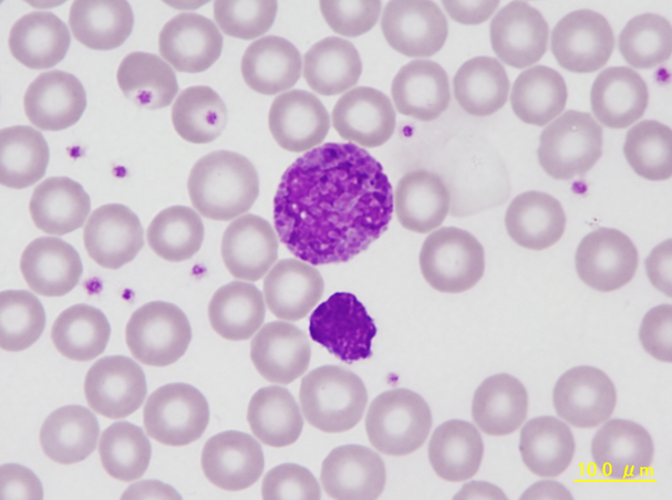
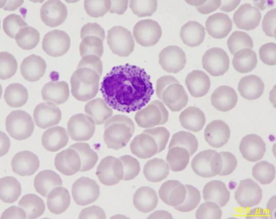
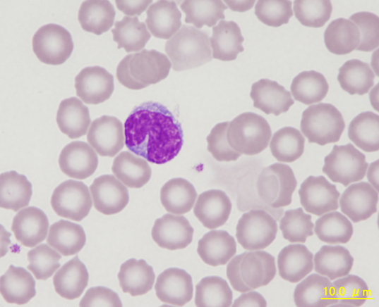
Basophil with intensely blue-purple and greyish blue granules, partially superimposing the nucleus. Below there is a bare nucleus of a lymphocyte.
<p>Basophil with intensely blue-purple and greyish blue granules, partially superimposing the nucleus. Below there is a bare nucleus of a lymphocyte.</p>
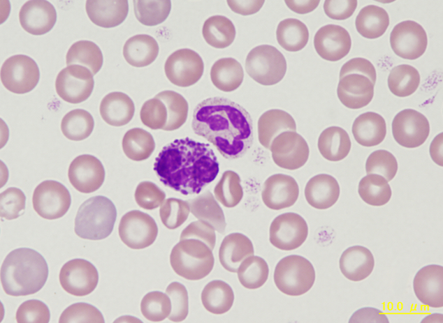
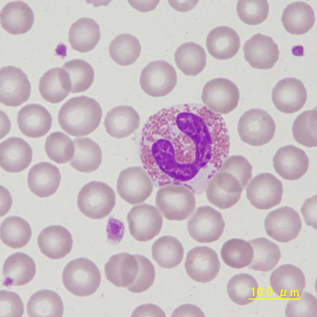
Blood smear of a marmoset showing a basophil with numerous, clearly visible purple staining granules next to a segmented neutrophil. Besides the commonly seen anisocytosis there are two large polychromatic red blood cells in the lower left.
<p>Blood smear of a marmoset showing a basophil with numerous, clearly visible purple staining granules next to a segmented neutrophil. Besides the commonly seen anisocytosis there are two large polychromatic red blood cells in the lower left.</p>
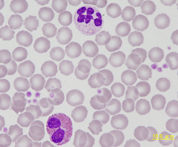
Eosinophils and neutrophils are somewhat harder to distinguish compared to other mammalian species due to their similar size and the presence of azurophilic granules in both cell types. Eosinophil granules are about double the size and rounder than those of neutrophils and show a brighter yellow-red colour. The cytoplasm of eosinophils is slightly more grey-blue and the
eosinophil nucleus is not as condensed or segmented.
<p>Eosinophils and neutrophils are somewhat harder to distinguish compared to other mammalian species due to their similar size and the presence of azurophilic granules in both cell types. Eosinophil granules are about double the size and rounder than those of neutrophils and show a brighter yellow-red colour. The cytoplasm of eosinophils is slightly more grey-blue and the </p> <p>eosinophil nucleus is not as condensed or segmented. </p>
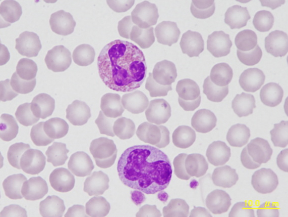
Eosinophil with red staining granules and poorly segmented nucleus at the top. Monocyte below showing the characteristic amorphous nucleus, greyish-blue cytoplasm and fine granules.
<p>Eosinophil with red staining granules and poorly segmented nucleus at the top. Monocyte below showing the characteristic amorphous nucleus, greyish-blue cytoplasm and fine granules.</p>
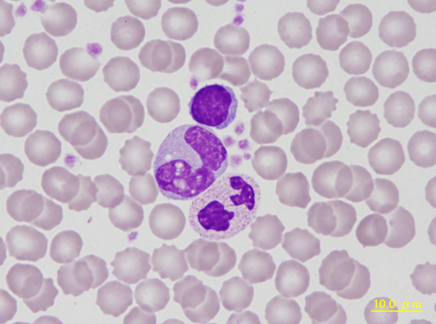
Typical pictures of a lymphocyte, monocyte and a neutrophil (top down). In the monocyte vacuoles overlap with the nucleus leading to the appearance of holes in the nucleus. The segments of the neutrophil nucleus are linked through nuclear filaments.
<p>Typical pictures of a lymphocyte, monocyte and a neutrophil (top down). In the monocyte vacuoles overlap with the nucleus leading to the appearance of holes in the nucleus. The segments of the neutrophil nucleus are linked through nuclear filaments.</p>
