Scientific Image Gallery
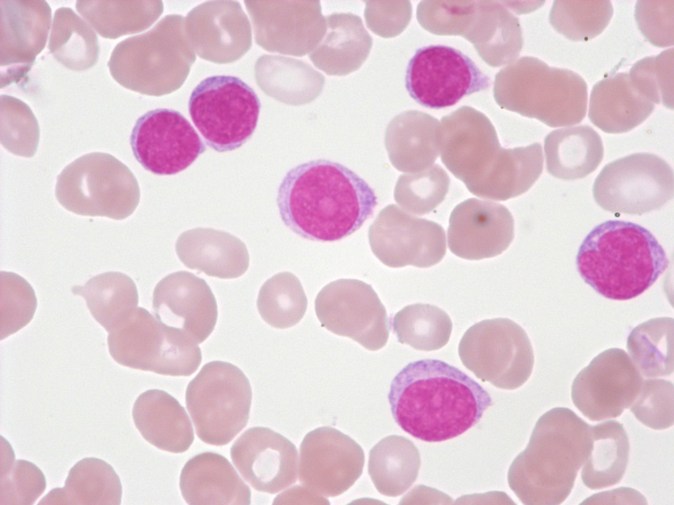
EDTA blood of a female patient with a transitional form of chronic lymphocytic leukaemia/prolymphocytic leukaemia. The three large cells are prolymphocytes, the smaller ones are lymphocytes.
<p>EDTA blood of a female patient with a transitional form of chronic lymphocytic leukaemia/prolymphocytic leukaemia. The three large cells are prolymphocytes, the smaller ones are lymphocytes.</p>
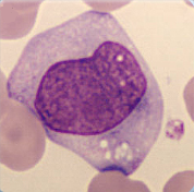
Cell description:
Size: bigger than monoblast
Nucleus: oval, kidney-shaped or lobulated, diffuse chromatin pattern, sometimes with nucleoli
Cytoplasm: pale basophil with fine azurophil granula.
Cell division is still possible.
<p>Cell description: </p> <p>Size: bigger than monoblast </p> <p>Nucleus: oval, kidney-shaped or lobulated, diffuse chromatin pattern, sometimes with nucleoli </p> <p>Cytoplasm: pale basophil with fine azurophil granula. </p> <p>Cell division is still possible.</p>
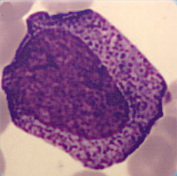
Cell description:
Size: 15-25 µm
Nucleus: oval with identifiable nucleoli and diffuse chromatin structure Cytoplasm: basophilic with visible golgi-zone and eye-catching azurophil granula (primary granulation)
<p>Cell description: </p> <p>Size: 15-25 µm </p> <p>Nucleus: oval with identifiable nucleoli and diffuse chromatin structure Cytoplasm: basophilic with visible golgi-zone and eye-catching azurophil granula (primary granulation)</p>
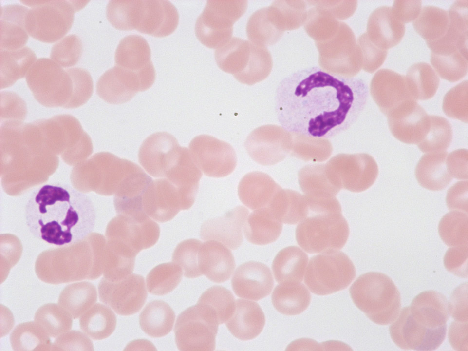
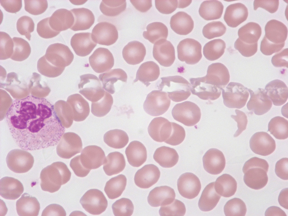
These so-called 'pseudo-Pelger's nuclear anomalies' with abnormal chromatin condensation and/or deficient nuclear segmentation are often found with viral infections, after chemotherapy, or with a myelodysplastic syndrome (MDS). Conversely, the true Pelger's nuclear anomaly is hereditary and without pathological significance.
<p>These so-called 'pseudo-Pelger's nuclear anomalies' with abnormal chromatin condensation and/or deficient nuclear segmentation are often found with viral infections, after chemotherapy, or with a myelodysplastic syndrome (MDS). Conversely, the true Pelger's nuclear anomaly is hereditary and without pathological significance.</p>
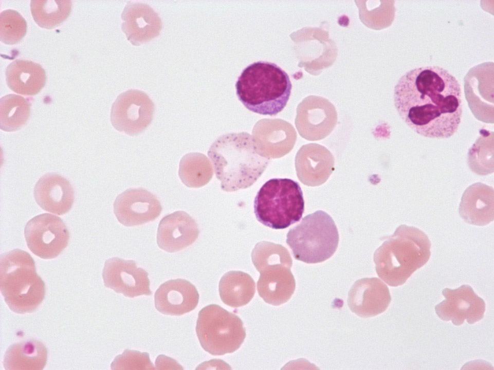
Large red blood cell with basophilic stippling (between the two lymphocytes) in a case of auto-immune haemolytic anaemia (AIHA). To the right, next to the lower lymphocyte, there is a polychromatic red blood cell.
<p>Large red blood cell with basophilic stippling (between the two lymphocytes) in a case of auto-immune haemolytic anaemia (AIHA). To the right, next to the lower lymphocyte, there is a polychromatic red blood cell.</p>
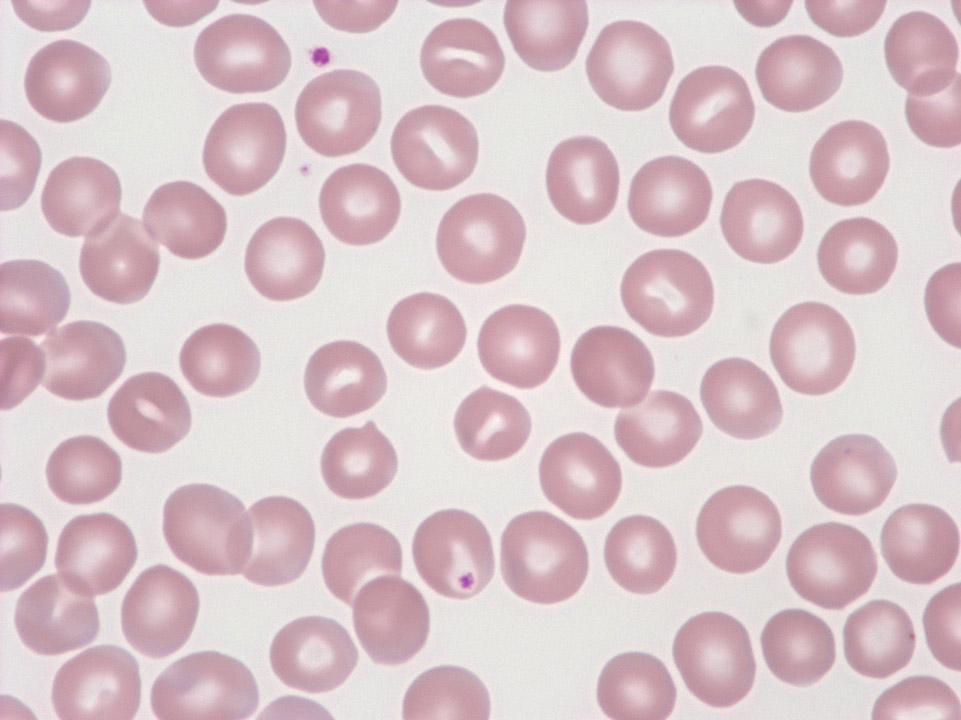
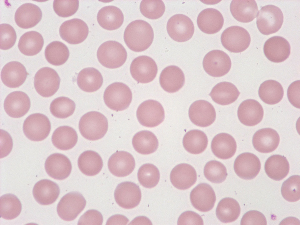
Red blood cells of a healthy individual showing a paler centre and a darker border. Occasionally the pale centre has an elongated shape (stomatocytes; at this frequency they have no pathological significance).
<p>Red blood cells of a healthy individual showing a paler centre and a darker border. Occasionally the pale centre has an elongated shape (stomatocytes; at this frequency they have no pathological significance).</p>
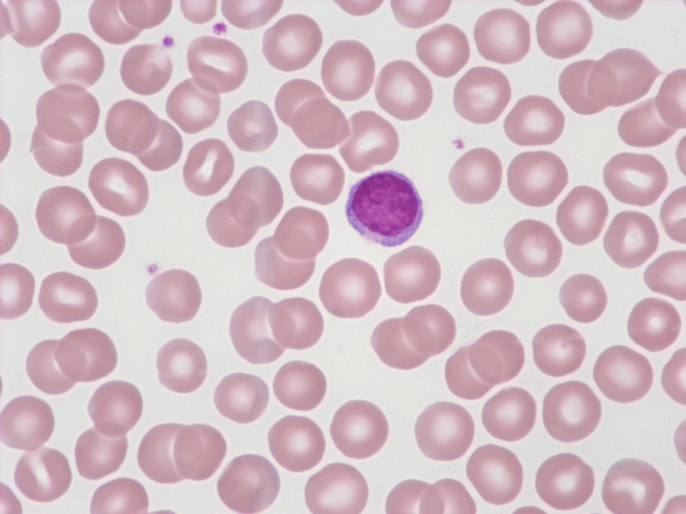
Red blood cells of a healthy individual. Normal red blood cells are a little smaller than the 'standard lymphocyte', which is pictured here.
<p>Red blood cells of a healthy individual. Normal red blood cells are a little smaller than the 'standard lymphocyte', which is pictured here.</p>
