Scientific Image Gallery
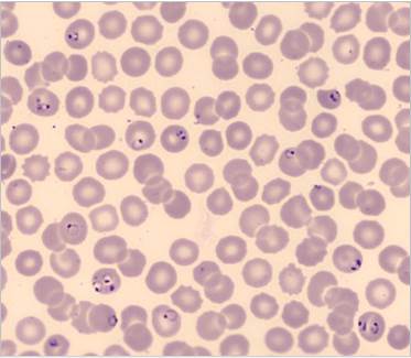
Trophozoites (rings) of Plasmodium falciparum are often thin and delicate, measuring on average 1/5 the diameter of the red blood cell. Rings may possess one or two chromatin dots. They may be found on the periphery of the RBC and multiple-infected RBC are not uncommon. Ring forms may become compact or pleomorphic, depending on the quality of the blood or if there is a delay in making smears.
<p>Trophozoites (rings) of Plasmodium falciparum are often thin and delicate, measuring on average 1/5 the diameter of the red blood cell. Rings may possess one or two chromatin dots. They may be found on the periphery of the RBC and multiple-infected RBC are not uncommon. Ring forms may become compact or pleomorphic, depending on the quality of the blood or if there is a delay in making smears. </p>
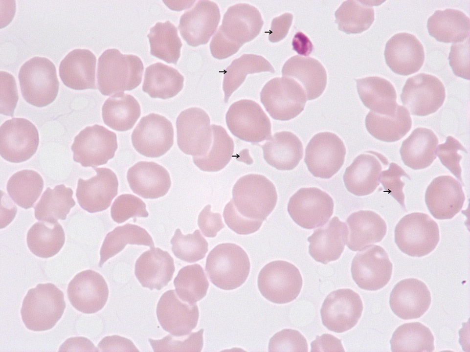
Schistocytes (->) and thrombocytopenia in a patient with haemolytic uraemic syndrome (HUS). This disease, as well as for example thrombotic thrombocytopenic purpura (TTP), is a so-called 'microangiopathic haemolytic anaemia' (MAHA). Characteristic of MAHA is a responsive increase in the production of red blood cells and platelets whose immature precursors (reticulocytes and immature platelet fraction, IPF) can be measured on certain Sysmex analysers. On a blood film the differential diagnosis of MAHA is not possible.
<p>Schistocytes (->) and thrombocytopenia in a patient with haemolytic uraemic syndrome (HUS). This disease, as well as for example thrombotic thrombocytopenic purpura (TTP), is a so-called 'microangiopathic haemolytic anaemia' (MAHA). Characteristic of MAHA is a responsive increase in the production of red blood cells and platelets whose immature precursors (reticulocytes and immature platelet fraction, IPF) can be measured on certain Sysmex analysers. On a blood film the differential diagnosis of MAHA is not possible.</p>
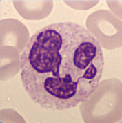
Cell description:
Size: 12-15 µm
Nucleus: clumped chromatin and mostly divided into 2-5 distinct segments connected with filaments
Cytoplasm: acidophilic with many fine reddish granules spread evenly Function: phagocytosis, play an important role in the unspecific immune defense, in the tissue they defend the mucosa against bacteria and fungi
<p>Cell description: </p> <p>Size: 12-15 µm </p> <p>Nucleus: clumped chromatin and mostly divided into 2-5 distinct segments connected with filaments </p> <p>Cytoplasm: acidophilic with many fine reddish granules spread evenly Function: phagocytosis, play an important role in the unspecific immune defense, in the tissue they defend the mucosa against bacteria and fungi</p>
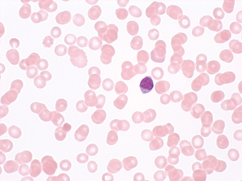
White blood cells 600/μL, granulocytes 10% (absolute: 60/μL), haemoglobin concentration 6 g/dL and platelets 10,000/μL. Diagnosis: severe aplastic anaemia. Monitoring before stem cell transplantation.
<p>White blood cells 600/μL, granulocytes 10% (absolute: 60/μL), haemoglobin concentration 6 g/dL and platelets 10,000/μL. Diagnosis: severe aplastic anaemia. Monitoring before stem cell transplantation.</p>
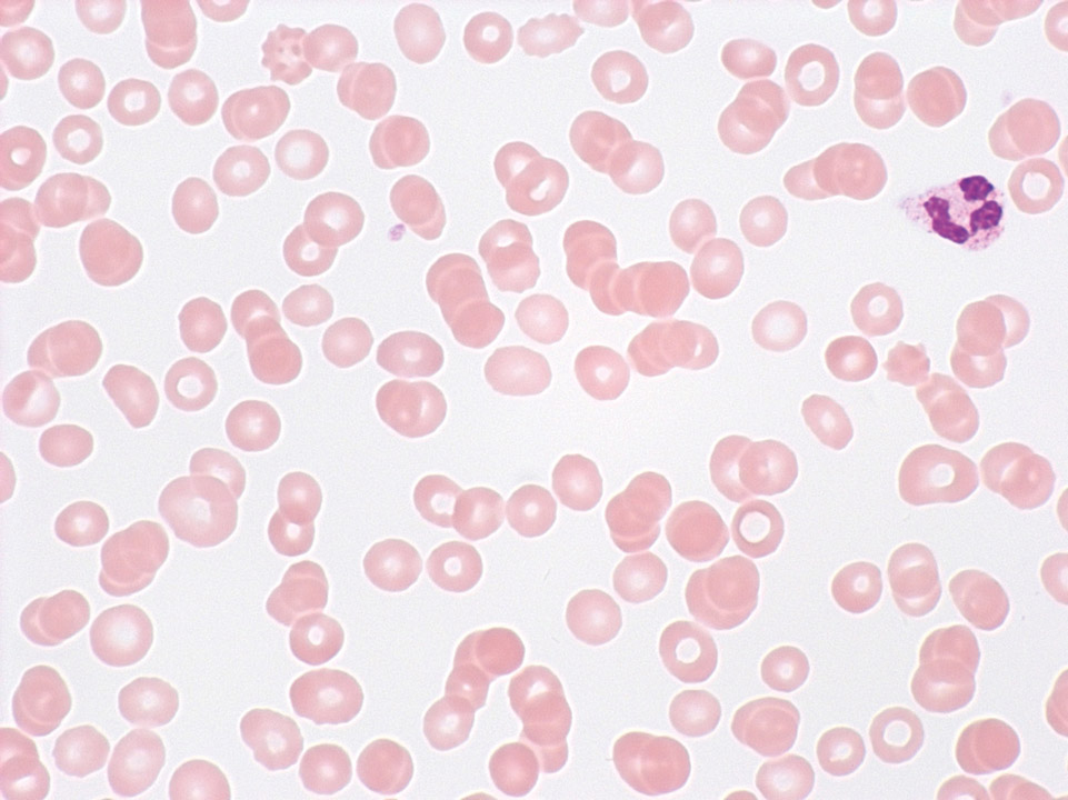
In severe anaemia (here severe aplastic anaemia with a haemoglobin concentration of 4.8 g/dL) or in erythrocytosis with a haematocrit above 50% it is difficult to prepare a proper blood film, with the blood film being either too thin (anaemia) or too thick. In cases of erythrocytosis it might be necessary to dilute the blood sample beforehand.
<p>In severe anaemia (here severe aplastic anaemia with a haemoglobin concentration of 4.8 g/dL) or in erythrocytosis with a haematocrit above 50% it is difficult to prepare a proper blood film, with the blood film being either too thin (anaemia) or too thick. In cases of erythrocytosis it might be necessary to dilute the blood sample beforehand.</p>
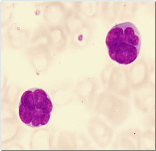
Typical for a Sézary cell is its cerebriform shape of the nucleus. The cytoplasm is usually basophilic with a medium nucleocytoplasmic ratio.
<p>Typical for a Sézary cell is its cerebriform shape of the nucleus. The cytoplasm is usually basophilic with a medium nucleocytoplasmic ratio. </p>
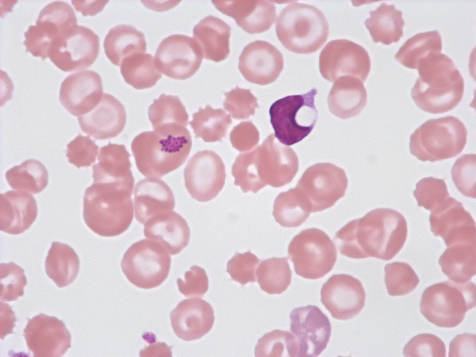
Accumulation of streptococci on a red blood cell of a patient in intensive care suffering from streptococcal septicaemia (further to the right, a lymphocyte with a cytoplasm vacuole can be seen).
<p>Accumulation of streptococci on a red blood cell of a patient in intensive care suffering from streptococcal septicaemia (further to the right, a lymphocyte with a cytoplasm vacuole can be seen).</p>
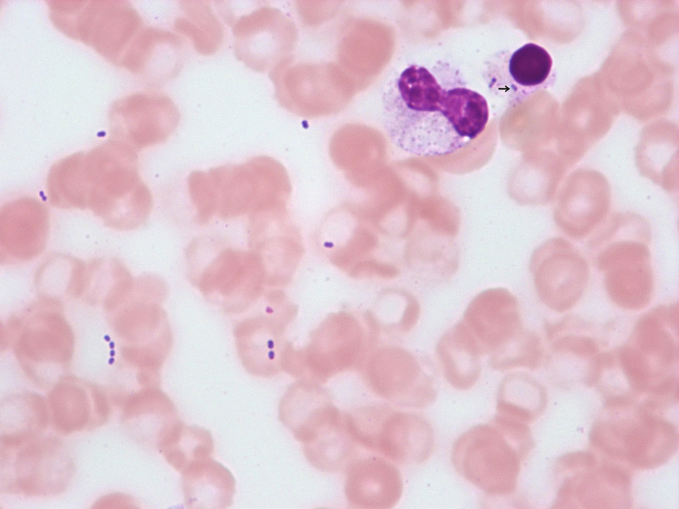
Streptococci, extracellular as well as intracellular (->), in a patient with finger gangrene. The patient died hours later despite intensive care treatment. If bacteria are detectable in a blood film, a blood film from a different patient should be stained with the same staining solution and be checked for bacteria to rule out contamination of the staining solution. If none are detectable, the detection of bacteria has to be reported to the treating physician immediately.
<p>Streptococci, extracellular as well as intracellular (->), in a patient with finger gangrene. The patient died hours later despite intensive care treatment. If bacteria are detectable in a blood film, a blood film from a different patient should be stained with the same staining solution and be checked for bacteria to rule out contamination of the staining solution. If none are detectable, the detection of bacteria has to be reported to the treating physician immediately.</p>
