Scientific Image Gallery

Bone marrow histology (Giemsa stain) with about 30% blast cells of a patient with AML. Two typical blasts are highlighted (->).
<p>Bone marrow histology (Giemsa stain) with about 30% blast cells of a patient with AML. Two typical blasts are highlighted (->).</p>
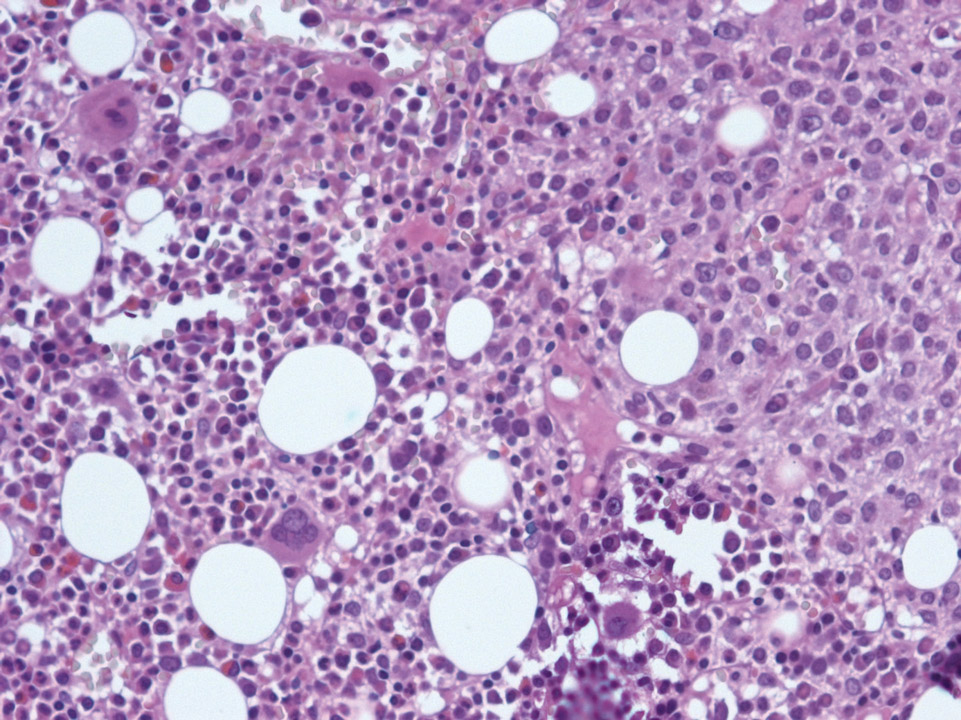
Bone marrow histology (haematoxylin eosin stain) with normal haematopoiesis on the left side. On the right side the bone marrow is infiltrated by a diffuse large B-cell lymphoma (DLBCL).
<p>Bone marrow histology (haematoxylin eosin stain) with normal haematopoiesis on the left side. On the right side the bone marrow is infiltrated by a diffuse large B-cell lymphoma (DLBCL).</p>

It is extremely rare that cancerous cells leak into the peripheral blood. Here, cells from a breast carcinoma are visible on the blood film.
<p>It is extremely rare that cancerous cells leak into the peripheral blood. Here, cells from a breast carcinoma are visible on the blood film.</p>

Cabot rings are round to oval or loop-like, fine, red-purple inclusions in red blood cells. They are remnants of the mitotic spindle apparatus. They can be seen with severe anaemia, e.g. thalassaemia, but are unspecific.
<p>Cabot rings are round to oval or loop-like, fine, red-purple inclusions in red blood cells. They are remnants of the mitotic spindle apparatus. They can be seen with severe anaemia, e.g. thalassaemia, but are unspecific. </p>
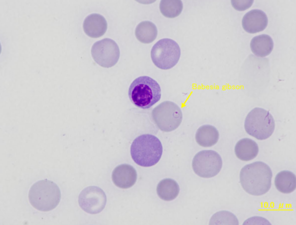
The yellow arrow points to a red blood cell infected with Babesia gibsoni, a protozoan transmitted by ticks. In the centre, there is a nucleated red blood cell. Overall, an increased number of polychromatic and differently sized red blood cells can be seen.
<p>The yellow arrow points to a red blood cell infected with Babesia gibsoni, a protozoan transmitted by ticks. In the centre, there is a nucleated red blood cell. Overall, an increased number of polychromatic and differently sized red blood cells can be seen. </p>
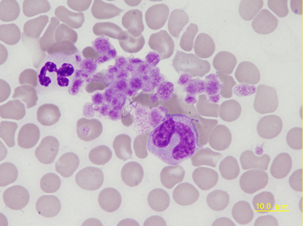
Aggregated large platelets, a segmented neutrophil (left) and a neutrophilic metamyelocyte (right). This sample was taken from a dog with chronic inflammation and acute haemorrhage a few days before.
<p>Aggregated large platelets, a segmented neutrophil (left) and a neutrophilic metamyelocyte (right). This sample was taken from a dog with chronic inflammation and acute haemorrhage a few days before. </p>
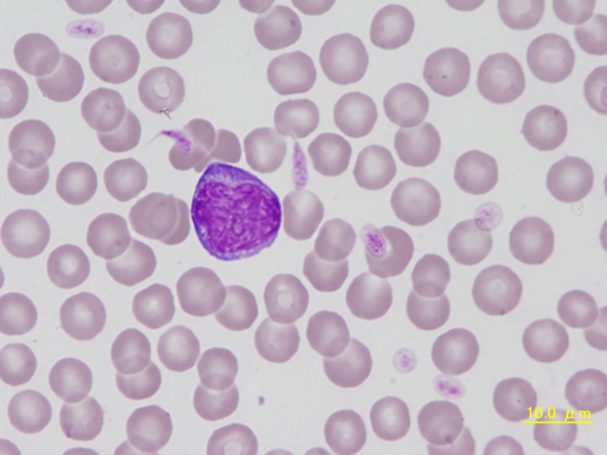
Atypical lymphocyte of a dog. The cell is large, with rough chromatin and dark blue cytoplasm. It is activated by various antigenic stimuli of the lymphatic tissue and changes morphologically.
<p>Atypical lymphocyte of a dog. The cell is large, with rough chromatin and dark blue cytoplasm. It is activated by various antigenic stimuli of the lymphatic tissue and changes morphologically.</p>
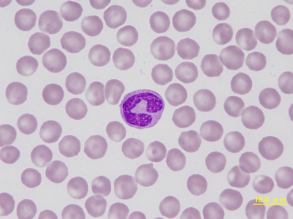
Band neutrophil with condensed chromatin and only mild constriction of the nucleus. The horseshoe shape of the nucleus is commonly seen with this young form of a neutrophilic granulocyte.
<p>Band neutrophil with condensed chromatin and only mild constriction of the nucleus. The horseshoe shape of the nucleus is commonly seen with this young form of a neutrophilic granulocyte.</p>
